Videos
March 2nd, 2014
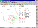
Digitising Demo
This video starts from scratch and finishes with a tracing.
The steps shown are:
- Create a patient folder.
- Create an x-ray montage.
- Import a cephalometric radiograph into the montage by dragging and dropping from Windows Explorer.
- Open the ceph. in Opal Image Viewer.
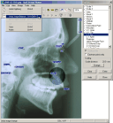
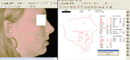
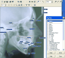
- Digitise using Opal Image Viewer.
- Save the ceph. back (storing the digitised points with it).
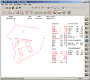
- Open the automatically generated digitising in Opal Tracing to complete it.
Morphing Demo
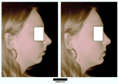
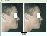
This video demonstrates how to create a prediction montage from a profile photo and a tracing.
The steps shown are:
- Create a photo montage.
- Import a profile photo into the montage by dragging and dropping from Windows Explorer.
- Open the photo in Opal Image Viewer.
- Digitise points on the photo in preparation for superimposing the x-ray tracing on the photo.
- Save the photo with its digitised points.
- Open an x-ray tracing.
- Connect the tracing and the photo together by dragging and dropping the tracing's date label onto the photo.
- Do some interactive surgical prediction on the tracing (a mandibular ramus osteotomy).
- Notice that the photo changes to reflect the changes in the tracing.
- Click on the 'Save' button in Opal Tracing.
- Notice that because we have a tracing and associated photo open at the same time, a special wizard is displayed.
- The wizard guides the user through the process of saving both the modified tracing and the modified photo. It also automatically creates a prediction montage.
- Close the tracing and the photo windows.
- Double-click on the prediction montage's icon to display it.
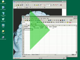
Area Measurement Demo
This video show how to measure areas and use them in a spreadsheet.
The steps shown are:
- Create a simple spreadsheet.
- Find a model montage to measure in Opal.
- Type patient name and date into the spreadsheet.
- Measure the area of a triangle and copy into the spreadsheet.
- Measure the area of an arch and copy into the spreadsheet.
- Calculate the ratio of areas.
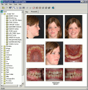
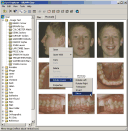
OPAL Screenshots
July 10th, 2010
-
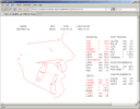
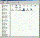
Some new Opal Explorer features in Opal 2.3.
-
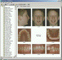
Some new Opal Image Viewer features in Opal 2.3.

-
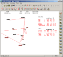
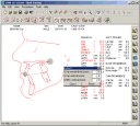
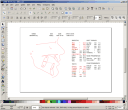
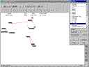
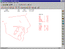
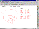
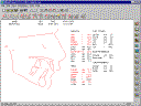
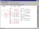
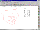
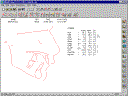
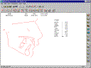
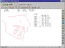
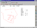
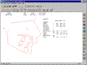
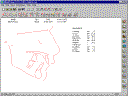
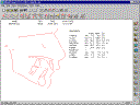
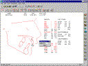
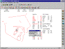
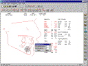
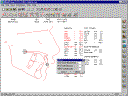
Some new Opal Tracing features in Opal 2.3.
-
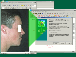
Image morphing.
-
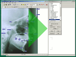
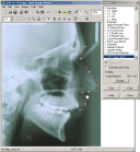
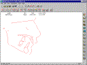
On-screen digitising.
-
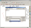
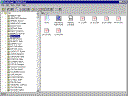
Full graphical interface for file creation and manipulation.
-
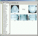
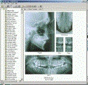
Image storage and presentation capabilities.
-
Digitising options: audio prompts, automatic magnification correction.


-
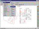
Multiple cephalometric analyses - Eastman, OPAL, Steiner, Downs, Ricketts,
McNamara.
-
Multiple display options.


-
Mouse manipulation for orthodontic and surgical procedures.
-
Drag-and-drop superimpositions.